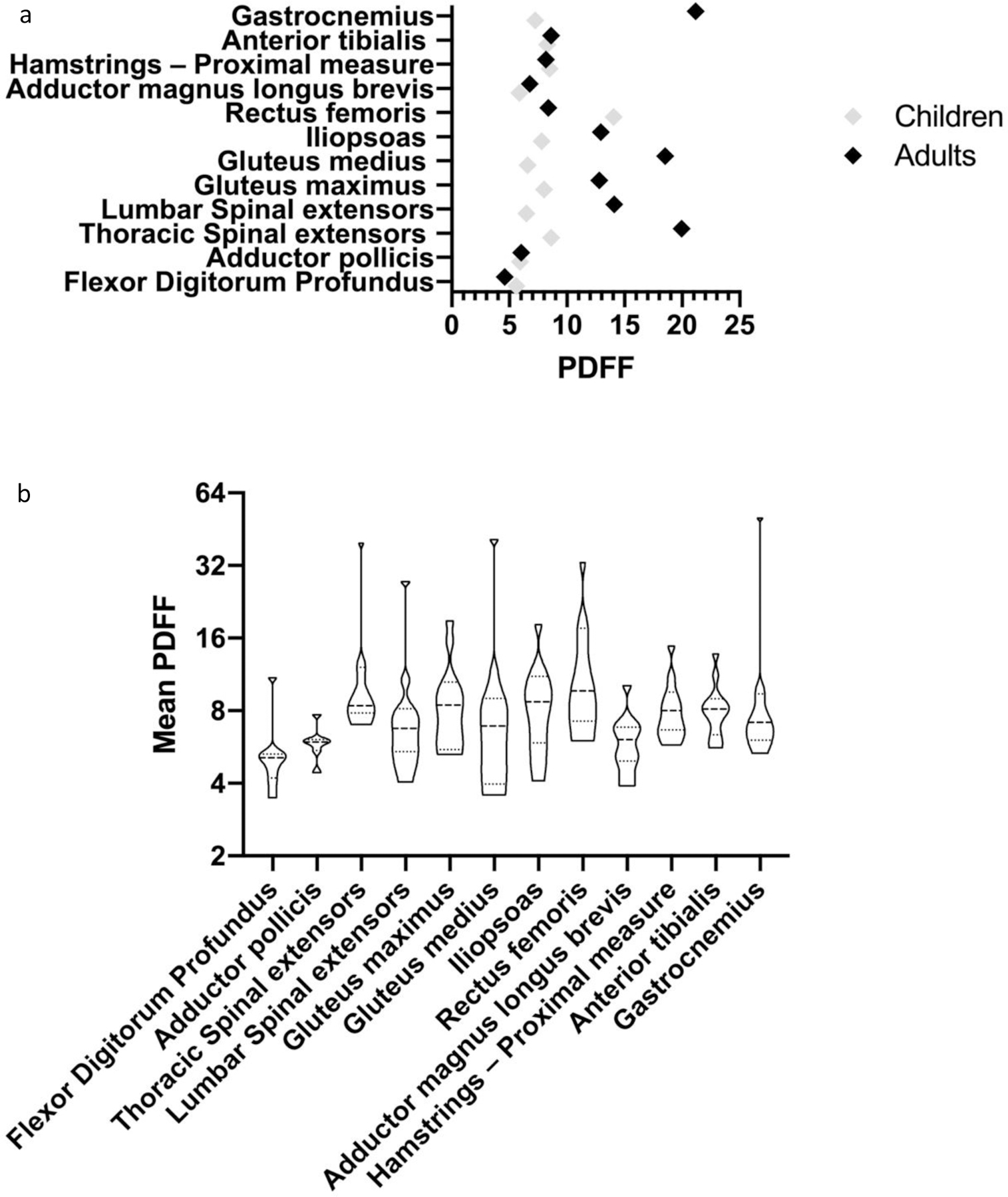
Figure 7: Mean PDFF for all muscles imaged divided by age group (a) and Mean PDFF for whole cohort (b).

Normal values for PDFF of muscles imaged falls between 2–5% for each muscle12–13. As demonstrated in the figures above, all muscles examined in both children and adults, except for flexor digitorum profundus in adults demonstrated an increased PDFF value indicating increased glycogen and fat deposition within the muscle. As demonstrated in the figure, adults with GSD IIIa demonstrated marked elevations in PDFF in the majority of muscles examined, most notably in the gastrocnemius, gluteus medius, and thoracic spinal extensors. In children, the most notably elevation in PDFF was observed in the rectus femoris, with a median value of 10.05 (IQR: 10.3).
