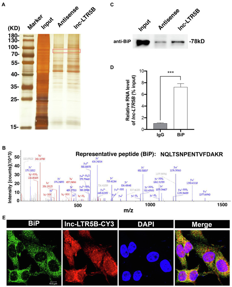
Figure 4.
Identification of BiP as a binding protein of lnc-LTR5B. (A) Silver staining of biotinylated lnc-LTR5B-associated proteins. The lnc-LTR5B-specific bands (highlighted bands) were excised and analyzed by mass spectrometry. (B) Mass spectrometry of highlighted band digests identifies the fragment ions from the representative peptide of BiP protein. (C) Western blot analysis of BiP from RNA pulldown assay using biotinylated lnc-LTR5B or antisense RNA. (D) RNA immunoprecipitation (RIP)-qPCR analysis of lnc-LTR5B immunoprecipitated by BiP antibody from DF-1 cells. Data are presented as the mean±SD, n=3; ***p<0.001 (two-tailed unpaired Student’s t test). (E) RNA FISH detecting endogenous lnc-LTR5B (red) combined with immunofluorescence staining of BiP (green) in DF-1 cells. DAPI staining is shown in blue. Scale bars, 10μm.

