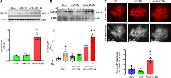
FIGURE 10.
MiR-18a-5p targets HIF-1α and mitochondrial fission. (A) Western blot and quantification showing expression of HIF-1α normalized to Actin in the H9c2 cardiomyocytes treated with scm, miR-18a-5p, and anti-miR-18a-5p. (B) Western blot and quantification showing expression of HIF-1α normalized to Actin in H9c2 cardiomyocytes treated by scm, miR-18a-5p, and anti-miR-18a-5p, with NE or no NE. Values are mean ± SEM, *P < 0.05 vs. scm, **P < 0.05 vs. miR-18a-5p. One-way ANOVA. (C) Top, live cell pDsRed2-Mito image of H9c2 cardiomyocytes treated with scm, miR-18a-5p, and anti-miR-18a-5p. Bottom, quantification of % number of cells with fragmented mitochondria (all globular) per unit areas in each group from six independent experiments.

