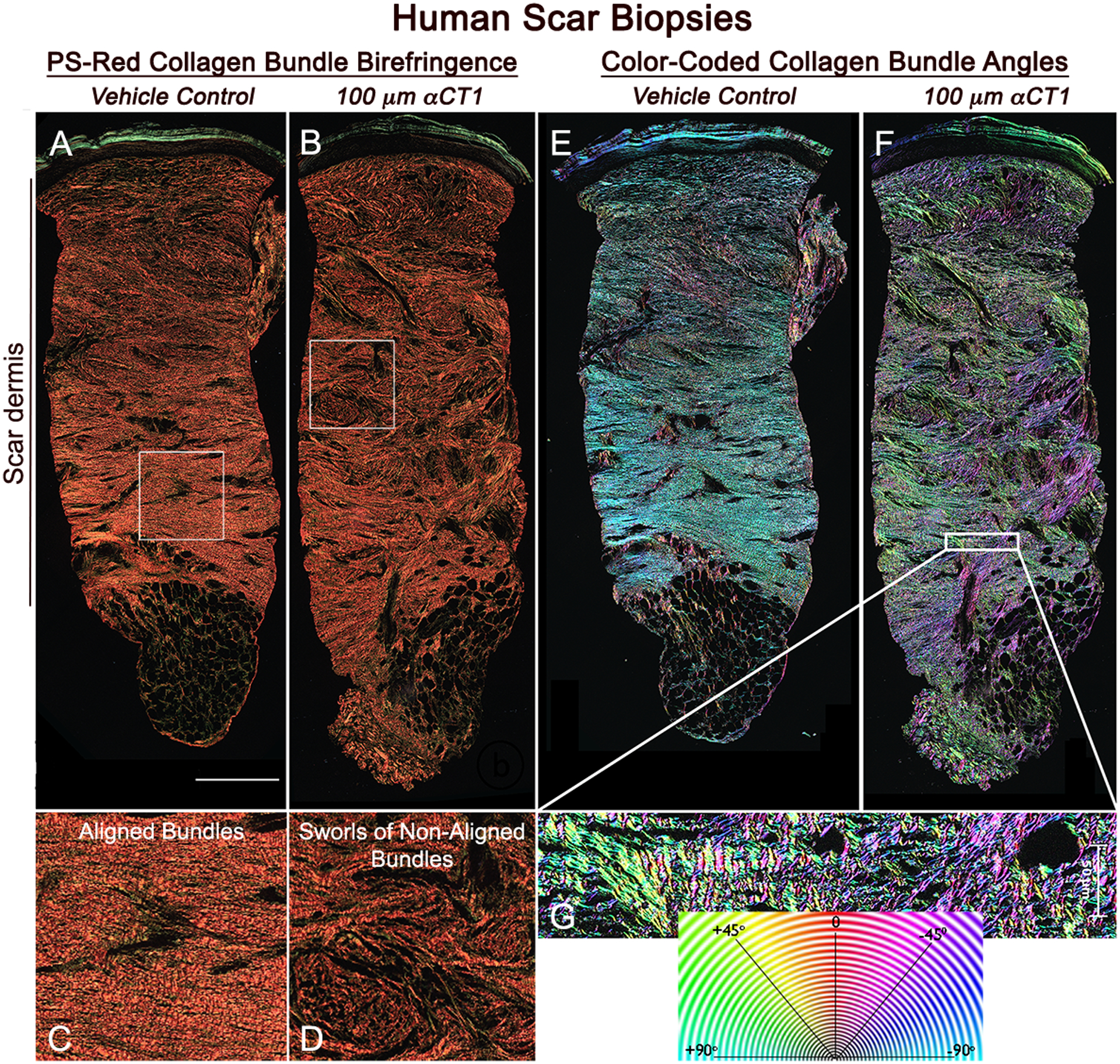
Figure 3. αCT1 effects on collagen organization in the human 29-day scar biopsies.

A) Olympus VS120 scan (20x objective) of a vehicle-control scar biopsied from the left arm of an individual in the patient group receiving the 100 μM therapeutic dose of αCT1. B) Corresponding scan of collagen birefringence of the αCT1 treated scar on the right arm of the same patient. C and D) Higher magnification views of the organization of collagen bundles in the boxed regions of (A) and (B), respectively. E and F) Collagen bundle angles in full-width sections of scars shown in (A) and (B) are color-coded over 180 degrees using OrientationJ software. H) Higher magnification views of boxed region in (F). Collagen bundles show a high degree of alignment in the control scar. In the αCT1-treated scar collagen bundles tend to be more randomly arranged and form larger sworls reminiscent of structures seen in unwounded skin. Scale = 0.5 mm
