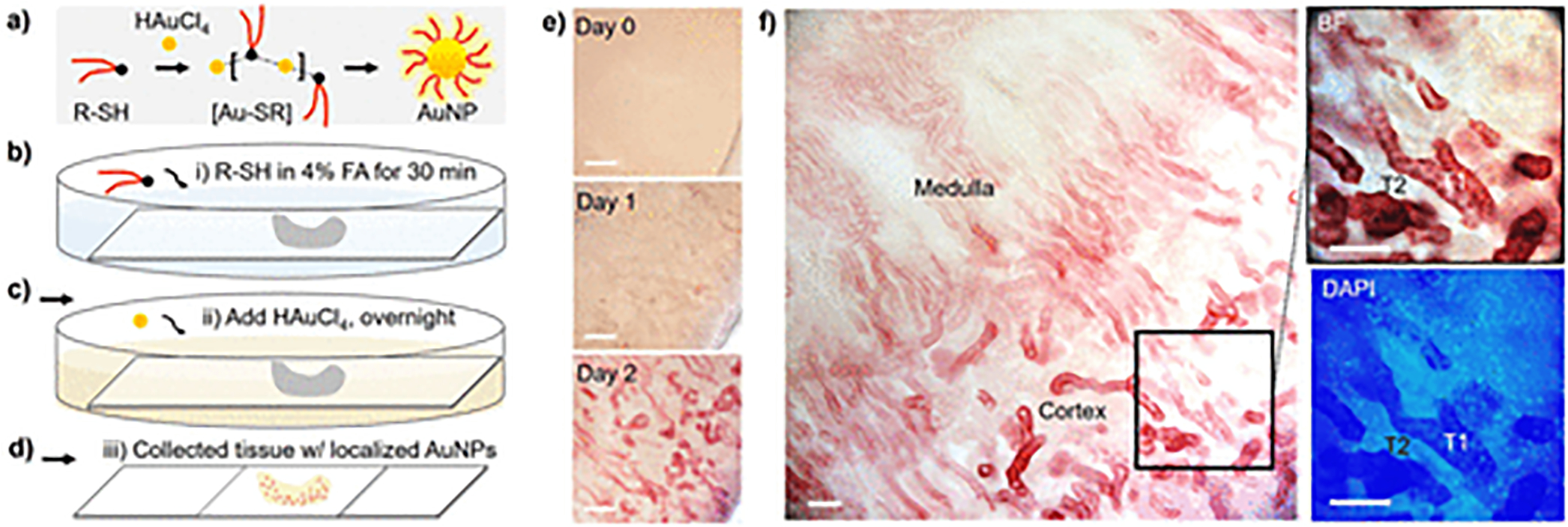
Figure 1.

In situ growth of AuNPs in biological (kidney) tissues. (a) Formation process of thiol-protected AuNPs in aqueous solution. The gold ions (HAuCl4) and thiol ligand (R-SH) in solution can readily form thiolated gold precursor ([Au-SR]) and then dissociate into thiol-protected AuNPs. (b–d) Scheme of in situ AuNP formation process in biological tissues (see Methods in SI for experimental details). FA, formaldehyde. (e,f) Time evolution of the AuNP growth in kidney tissues. The AuNPs (plasmonic, red color) showed preferential distribution in the selected tubules (high in T1, but low in T2, f) instead of other nephron segments in kidney cortex. Scale bar, 0.2 mm (e); 0.1 mm (f).
