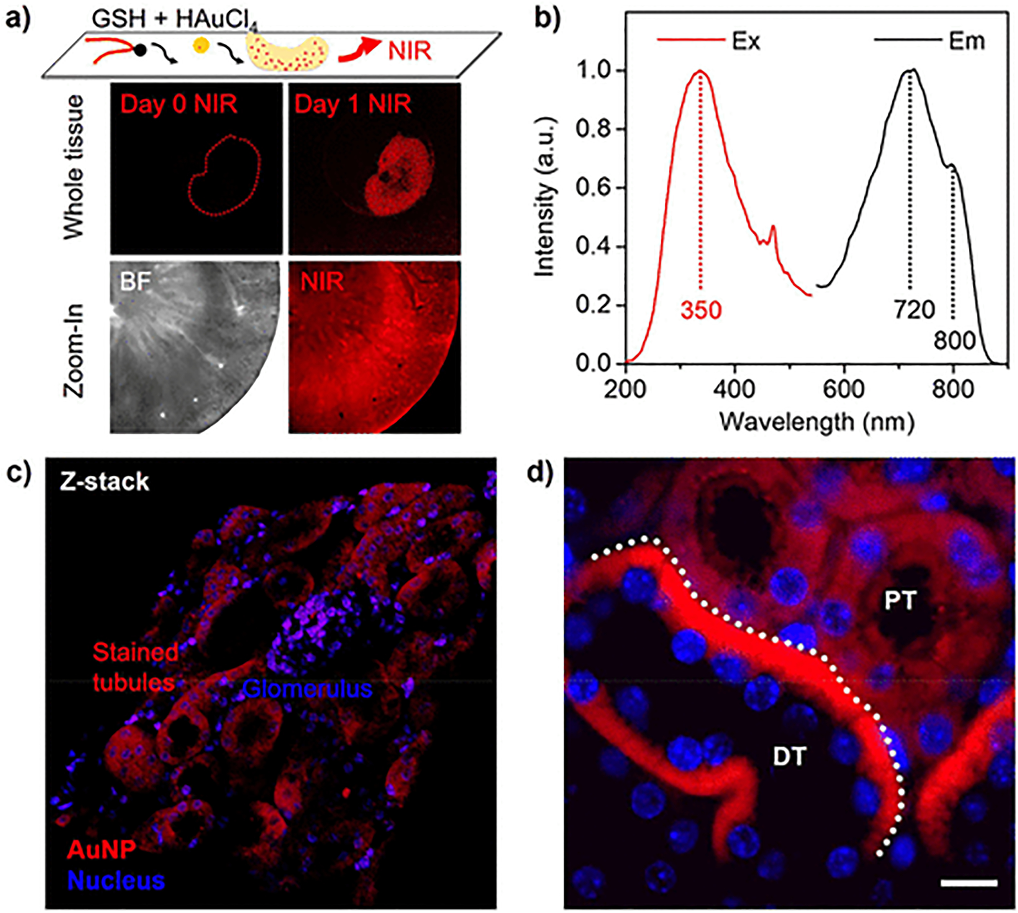
Figure 3.

GSH-directed growth of NIR luminescent AuNPs in kidney tissues. (a) Whole-tissue NIR fluorescence imaging of the kidney before (Day 0) and after (Day 1) the formation of GS-AuNPs and zoom-in images of bright field (BF) and near-infrared (NIR) imaging of the kidney cortex after the in situ AuNP growth. (b) NIR luminescence spectra of the GS-AuNPs. (c) Confocal fluorescence microscopy (z-stack) image of kidney cortex with localized GS-AuNPs (full-scale width, 320 μm). (d) Microscopy imaging for the kidney slides (4 μm) with in situ formed ultrasmall GS-AuNPs. Scale bar, 10 μm. DT, distal tubule; PT, proximal tubule.
