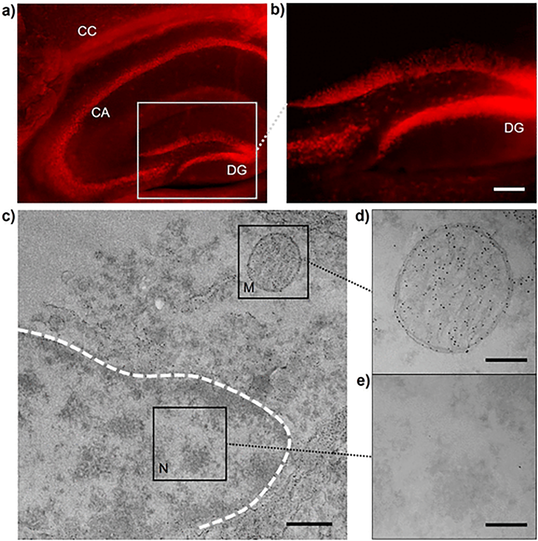
Figure 4.

GSH-directed growth of AuNPs in brain hippocampus. (a,b) NIR fluorescent microscopy imaging of GS-AuNPs in brain hippocampus. Scale bar, 100 μm. (c–e) TEM imaging of subcellular area of hippocampus cells with GS-AuNPs growth. M, mitochondrion; N, nucleus. Scale bar, 500 nm (c); 200 nm (d,e).
