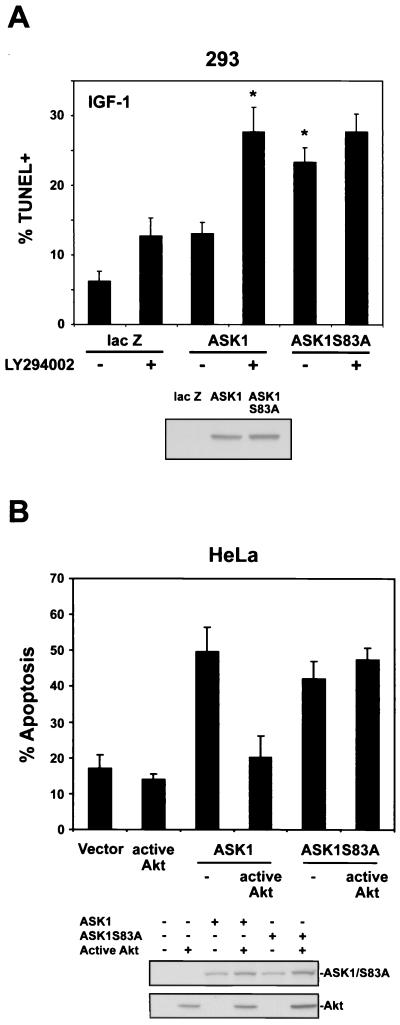
FIG. 5.
Activation of the PI3-K/Akt pathway decreases ASK1-mediated apoptosis. (A) 293 cells were cotransfected with 1.75 μg of lacZ or HA-tagged ASK1 constructs with 0.1 μg of pDsRed as a marker for 24 h. Cells were incubated with or without LY294002 (10 μM) for 1 h, and then all cells were exposed to 10 ng of IGF-1/ml for 36 h. Cell death was assessed by TUNEL-FITC staining and flow cytometry for both TUNEL positivity and DsRed positivity to assay transfected cells only (upper panel). All bars represent means plus SEM from four to seven independent experiments. The asterisk indicates a significant difference from ASK1 at P < 0.05 by one-way ANOVA followed by the Bonferroni t test. In parallel, sister cultures were transfected with solutions identical to those used in cultures later assessed for cell death. Cells were lysed in 1% NP-40 buffer after 24 h, and lysates were probed for expression of ASK1 constructs with anti-HA (bottom panel). (B) HeLa cells were cotransfected with 0.75 μg of the indicated ASK1 constructs plus 0.25 μg of EGFP-IRES-HA-Akt (E40K) (active Akt) or EGFP-IRES (Vector) for 24 h and then serum starved for 24 h. Cells were labeled with Hoechst 33342, and EGFP-positive cells were scored for apoptosis by nuclear morphology. In each experiment, 100 EGFP-positive cells were counted per condition. The histogram represents means plus SEM from four independent experiments. As described for panel A, sister cultures were transfected with solutions identical to those used in cultures assessed for cell death. Cell lysates were probed for expression of ASK1 constructs and active Akt with anti-HA (bottom panel).

