. 2021 Dec 2;10:e71656. doi: 10.7554/eLife.71656

Figure 3. The interaction of CGI-99 and FAM98B is mediated via their N-terminal regions.

(A) Crystal structure of the N-terminal region of human CGI-99 (residues 2–101). The N- and C-termini, as well as the N-terminal extension of CGI-99 (residues 2–18), are indicated. (B) DALI pairwise alignment of CGI-99(2-101) (yellow) with human NDC80 (blue, PDB ID: 2ve7). (C) Pull-down assays using pre-immobilized GST-CGI-99(2-101) with lysates from HEK293T cells transiently overexpressing HA-(StrepII)2-GFP-FAM98B constructs. The input (I) and bound (B) fractions were analyzed by SDS-PAGE and visualized by in-gel GFP fluorescence (upper panel) followed by Coomassie Blue staining (lower panel). The ‘no bait’ control contained HEK293T lysate incubated with GSH resin in the absence of GST-CGI-99, ‘no prey ctrl’ contained GSH resin incubated with GST-CGI-99(2-101).

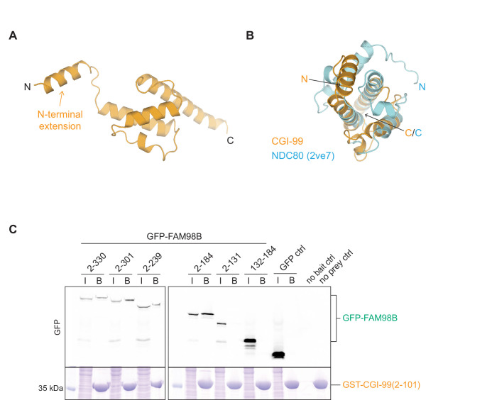
Figure 3.

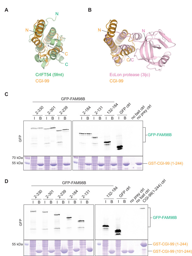
Figure 3—figure supplement 1. Analysis of the N-terminal domain of CGI-99 and the CGI-99:FAM98B interaction.

Figure 3—figure supplement 1.

(A, B) DALI structural alignment of the N-terminal domain of CGI-99 (yellow) with (A) Chlamydomonas reinhardtii IFT54 (green, PDB ID: 5fmt) and (B) Escherichia coli Lon protease (pink, PDB ID: 3ljc). N- and C-termini are indicated. (C, D) Pull-down assays of immobilized (C) GST-CGI-99(1-244) or (D) GST-CGI-99(101-244) with lysates of HEK293T cells transiently overexpressing HA-(StrepII)2-GFP-FAM98B constructs. The input (I) and bound (B) fractions were analyzed by SDS-PAGE and visualized by in-gel GFP fluorescence (upper panel) followed by Coomassie Blue staining (lower panel). The ‘no bait’ control contained HEK293T lysate incubated with GSH resin in the absence of GST-CGI-99, ‘no prey ctrl’ contained GSH resin incubated with one of the GST-CGI-99 constructs, and ‘CGI-99(1-244) control’ is the bound fraction of GSH-immobilized GST-CGI-99(1-244) incubated with HEK293T cell lysate expressing HA-(StrepII)2-GFP-FAM98B(2-301).