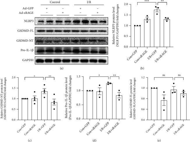
Figure 5.

Effects of sRAGE on the expression of pyroptosis-associated proteins in primary cultured cardiomyocytes following ischemia-reperfusion injury. I/R groups were subjected to ischemia for 2 hours and reperfusion for 24 hours. (a) Representative images of Western blot for pyroptosis-associated proteins. The histogram depicts the quantitative densitometry analysis of Western blot data. (b) Western blot analysis of NLRP3 protein expression. (c) Western blot analysis of the NH2-terminal cleaved GSDMD protein level. (d) Western blot analysis of the pro-IL-1β protein level. (e) Western blot analysis of the full-length GSDMD protein level. GAPDH was used as the internal reference control. Data are expressed as mean ± SEM (n ≥ 3 replicates). ∗p < 0.05; ∗∗p < 0.01; ∗∗∗p < 0.001; ns: no significance.
