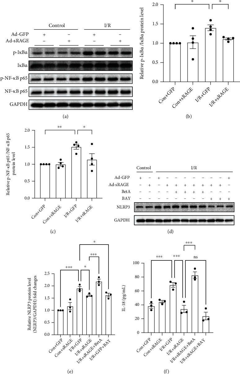
Figure 6.

Effects of sRAGE on the NF-κB pathway in primary cardiomyocytes against I/R-induced pyroptosis. (a) Representative images of Western blot for the phosphorylation level of IκBα and NF-κB p65 proteins. The histogram depicts the quantitative densitometry analysis of Western blot data. (b) Western blot analysis of the phosphorylation level of IκBα. (c) Western blot analysis of the phosphorylation level of NF-κB p65. GAPDH was used as the loading control. (d) Representative images of Western blot for the NLRP3 level. I/R groups were subjected to ischemia for 2 hours and reperfusion for 24 hours. (e) Western blot analysis of the NLRP3 level. (f) The level of IL-18 cytokines excreted by primary cultured cardiomyocytes within 24 hours of reperfusion. Data are expressed as mean ± SEM (n ≥ 3 replicates). BetA: betulinic acid; BAY: BAY117082; ∗p < 0.05; ∗∗p < 0.01; ∗∗∗p < 0.001; ns: no significance.
