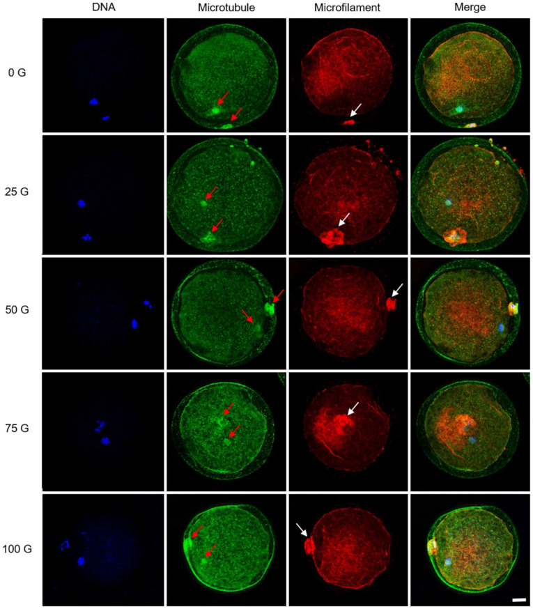
Fig. 4.
Confocal microscopy of cytoskeleton distribution in mature porcine oocytes. The microtubules were distributed in the meiotic spindle of the oocytes and the first polar body (red arrows). The expression of microfilaments strongly localizes around the position of the first polar body (white arrows). No distinct alterations of these oocytes can be detected after treatment with various IF-EMFs. Blue: chromosomes; green: microtubules; red: microfilaments. Scale bar: 25 µm.

