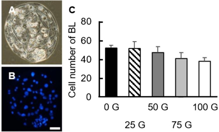
Fig. 7.
Total cell numbers of day 7 porcine blastocysts in the various IF-EMF treatment groups. (A, B) The representative micrographs of the blastocyst embryos in bright field (A) and DAPI staining (B). Blue: nuclei. Scale bar: 25 µm. (C) Average total cell numbers of the blastocysts (day 7) in each treatment group after parthenogenetic activation. No difference between any two groups can be detected (P > 0.05). BL: blastocyst.

