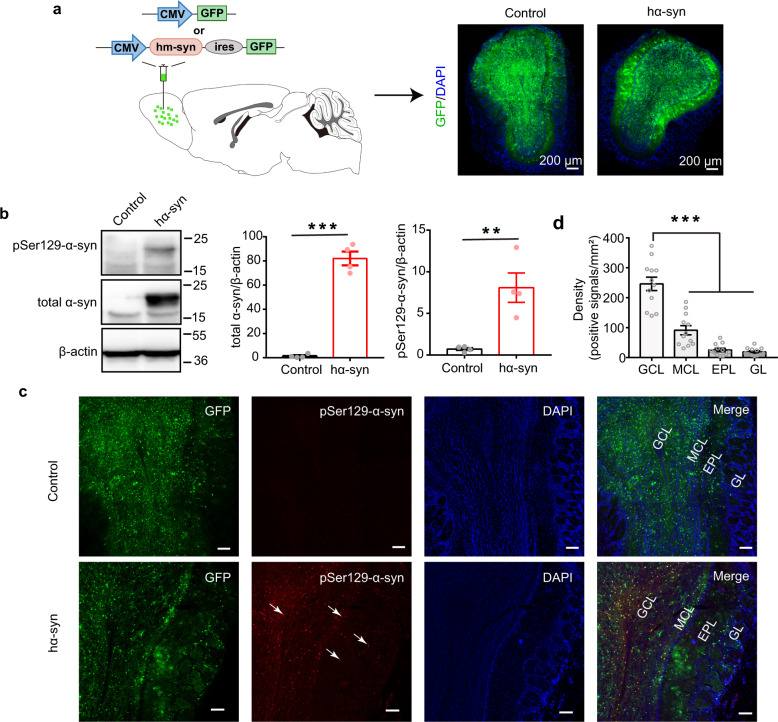
Fig. 1. Exogenous high human α-synuclein expression generates aggregates in the OB.
a Schematic of the viral injections (upper) and the GFP-labeling patterns in the OB of animals injected with AAV-CMV-hm-syn-ires-GFP (hα-syn) or AAV-CMV-GFP (control) (lower). b The expression of total α-syn and pSer129-α-syn in OB tissue 3 weeks after viral injection, measured with western blots. n = 4 mice for each group. All blots derive from the same experiment and that they were processed in parallel. c Representative images showing GFP-positive, pSer129-α-syn-positive, and co-labeled cells in different layers of the OB 3 weeks after viral injection. The white arrows indicate α-syn aggregates. Scale bars = 100 μm. d Quantitative analysis of the density of pSer129-α-syn-positive signals in different layers of OB. n = 12 slices from 4 mice for each group. GCL granule cell layer, MCL mitral cell layer, EPL external plexiform layer, GL glomerular layer, α-syn α-synuclein; **p < 0.01; ***p < 0.001. Data are presented as mean ± SEM.

