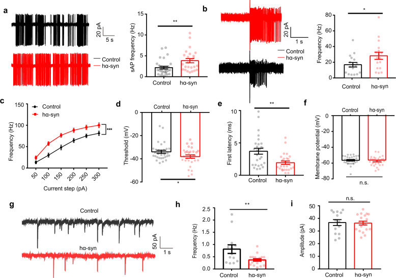
Fig. 5. α-Synuclein aggregates increase spontaneous and evoked firing in mitral cells in vitro.
a Representative (left) and quantitative analysis (right) of spontaneous firing rates of mitral cells (control: 30 cells from 13 mice; hα-syn: 25 cells from 15 mice) over a period of 5 min. b Representative (left) and quantitative analysis (right) of olfactory-nerve-evoked firing rates of mitral cells (17 cells from 11 control mice and 17 cells from 8 hα-syn group mice) in a 1.8 s recording period (olfactory nerve stimulation; 200 ms, 400 mA). c–f Quantitative analysis of the frequency (c), threshold voltage to trigger first AP (d), onset latency (e), and membrane potential (f) elicited by positive current injections (control: 30 cells from 11 mice; hα-syn: 29 cells from 7 mice). g Representative mIPSCs recorded from M/Ts in control and hα-syn slices. h, i Quantitative analyses of mIPSCs frequency (h) and amplitude (i) over a 5-min recording period (n = 14 neurons from 5 mice and 19 neurons from 4 mice; control and hα-syn groups, respectively). sAP spontaneous action potential, mIPSC miniature inhibitory postsynaptic current; *p < 0.05; **p < 0.01; ***p < 0.001; n.s. not significant. Data are presented as mean ± SEM.

