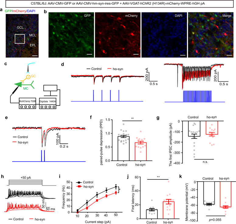
Fig. 6. α-Synuclein aggregates reduce paired-pulse depression of light-evoked IPSCs recorded from mitral cells and decrease granule cell activity in the OB.
a, b Low-magnification (scale bars = 100 μm) (a) and high-magnification (scale bars = 20 μm) (b) images showing granule cells labeled with mCherry in the OB. EPL external plexiform layer, MCL mitral cell layer, GCL granule cell layer. c Schematic of the light-evoked IPSC electrophysiological recording. d Representative IPSCs recorded from mitral cells when 2 Hz (left) and 20 Hz (right) light illumination was applied. e Representative traces of paired-pulse IPSCs. f Quantitative analysis of the paired-pulse depression of light-evoked IPSCs recorded from mitral cells from the control and hα-syn groups (control: 19 cells from 8 mice; hα-syn: 14 cells from 6 mice). g Quantitative analysis of the amplitude of the first light-evoked IPSC. h Representative current injection-evoked APs in granule cells. i–k Quantitative analysis of the frequency (i), onset latency (j), and membrane potential (k) elicited by positive current injections (13 cells from 5 mice for the control and hα-syn groups, respectively). mIPSC miniature inhibitory postsynaptic current; *p < 0.05; **p < 0.01; n.s. not significant. Data are presented as mean ± SEM.

