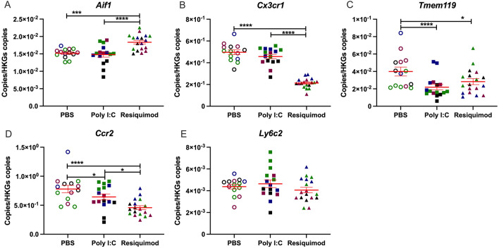
Figure 4.
Effect of MIA by poly I:C or resiquimod on microglial markers in foetal brain. Foetal brain tissues were collected after 4 h MIA (PBS, poly I:C (20 mg/kg, LMW), resiquimod (2 mg/kg)). (A,B) Aif1 was increased by resiquimod, however, Cx3cr1 was downregulated by resiquimod compared to PBS. Both genes were not changed by poly I:C. (C) Tmem119 was significantly downregulated by MIA compared to PBS. (D, E) Ccr2 was significantly downregulated by MIA compared to PBS, on the other hand, Ly6c2 was not changed by MIA. Absolute quantification was performed via RT-qPCR and the data were normalised to Gapdh and Tbp. The individual dots are shown along with mean ± SEM. Colour indicates dams within a single treatment (same colour means “same dam”). The data were log transformed and analysed by two way-ANOVA, Tukey post-hoc test (n = 14–18 independent samples; *p ≤ 0.05, **p ≤ 0.005, ***p ≤ 0.001, ****p ≤ 0.0001). Details of ANOVA F values and p values are provided in Supplementary Table 1.

