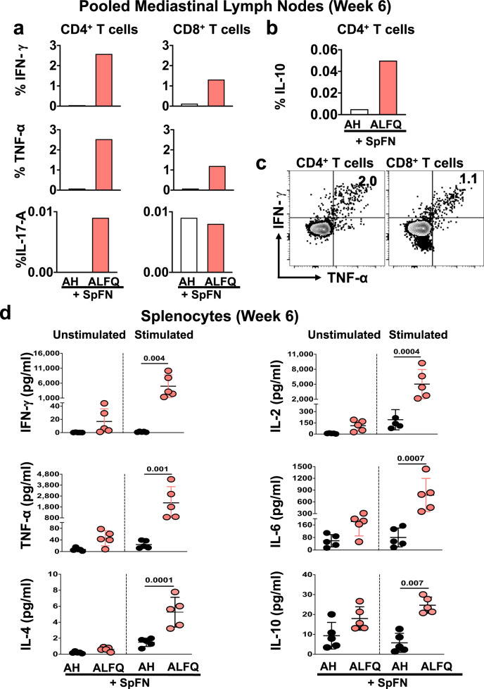
Fig. 4. Cytokine responses in the pooled mediastinal lymph nodes and spleens of mice after SpFN prime-boost vaccination.
a, b Percentage of SARS-CoV-2 spike-specific T cells in the mediastinal lymph nodes at week 6 (3 weeks post-prime-boost vaccination). a Percentage of CD4+ and CD8+ T cells expressing IFN-γ, TNF-α, IL-17A, and b CD4+ T cells expressing IL-10. Each sample is representative of the pooled lymph nodes within the vaccine group. c SARS-CoV-2 spike-specific CD4+ and CD8+ T cells in the mediastinal lymph nodes following prime-boost vaccination with SpFN + ALFQ co-express IFN-γ and TNF-α upon peptide stimulation. d Splenocytes were stimulated with SARS-CoV-2 spike-specific peptides and cytokines in the culture supernatants were measured by the ECLIA-based multiplex MSD platform. Dots represent data from individual mice (n = 5/group), horizontal lines represent mean and error bars represent s.d. Differences between the two groups were analyzed by using a nonparametric Mann–Whitney U-test with p ≤ 0.05 considered as statistically significant.

