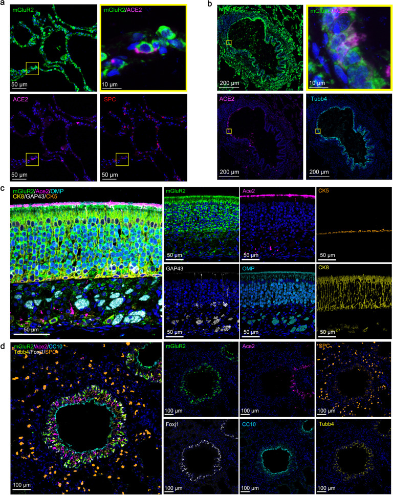
Fig. 4. mGluR2 is expressed in the respiratory system.
a, b Multiplex immunofluorescence staining for the detection of mGluR2-positive cells in a normal human lung section. mGluR2 (green), ACE2 (magenta), Tubb4 (cyan), and SPC (red). The yellow areas are shown adjacently at a higher magnification in the alveoli (a) or bronchia (b). c Multiplex immunofluorescence staining for the detection of mGluR2-positive cells in olfactory epithelium sections of young mouse. mGluR2 (green), Ace2 (magenta), GAP43 (white), CK5 (gold), CK8 (yellow), and OMP (cyan). d Multiplex immunofluorescence staining for the detection of mGluR2-positive cells in lung sections of young mouse. mGluR2 (green), Ace2 (magenta), Foxj1 (white), SPC (gold), Tubb4 (yellow), and CC10 (cyan).

