Figure 6.
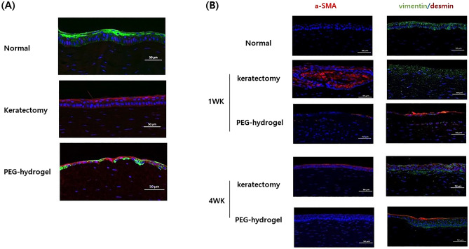
Fluorescent confocal microscopy images of rabbit keratectomy cornea without treatment and with polyethylene glycol (PEG)-collagen hydrogel application. (A) Cy5 stained with PEG-collagen hydrogel (magenta) was maintained underneath a multilayered, migrated epithelial layer stained with CK3 (green) after 1 week of keratectomy. (B) Immunohistochemical staining of α-smooth muscle actin (α-SMA), vimentin, and desmin markers for myofibroblasts in the keratectomy-subjected rabbit cornea over 1 week and 4 weeks. Note the high density of α-SMA (red) and vimentin (green) in the anterior stroma of rabbit corneas at 1 week after keratectomy alone. After 4 weeks, α-SMA (red) expression remained in the epithelium in the keratectomy-only group, whereas low α-SMA expression was found in the keratectomy with PEG-collagen hydrogel-treated group. Desmin (red) was expressed in the epithelium of the keratectomy with PEG-collagen hydrogel-treated group after 1 to 4 weeks.

