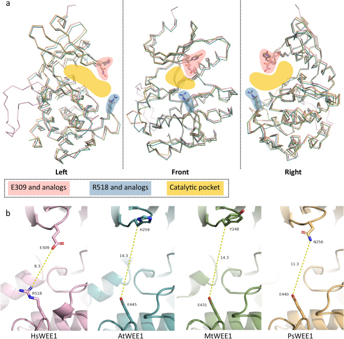
Figure 5.
Access to the catalytic pocket of WEE1. (a) Front and side views of H. sapiens WEE1 (pink) 3D structure superimposed to A. thaliana (blue), M. truncatula (green) and P. sativum (gold) predictions in ribbon representation. Residues conditioning the access to the catalytic pocket are highlighted in pink for E309 and its analogs and in blue for R518 and its analogs. The resulting catalytic pocket space approximately occupied by the target is highlighted in yellow. (b) Distance measurement (yellow hatched lines) in Å between residues conditioning the access to the catalytic pocket for the four 3D structures.

