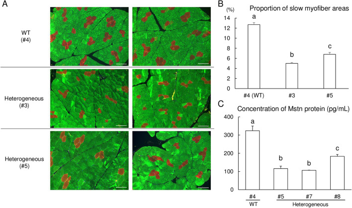
Figure 6.
Immunohistochemical assessment and quantification of MSTN protein concentration of wild-type (WT) and MSTN heterogeneous mutant piglets. (A) The longissimus dorsi muscles biopsies derived from WT (#4) and mutant piglets (#3 and #5) were immunohistochemically stained for slow (red) and fast (green) skeletal muscle myosin. The scale bar in each panel represents 100 μm. (B) Proportion of slow myofibers in longissimus dorsi muscle tissues. The slow myofiber areas were calculated as percentages from seven images after immunofluorescence staining for slow and fast type muscle fiber markers in longissimus dorsi muscle tissues obtained from 40-day-old piglets. WT, wild-type. Each bar represents a mean ± SEM. a–cp < 0.05. (C) Comparison of MSTN protein concentrations. Equal concentrations (1.0 mg mL−1) of total protein extracts obtained from the longissimus dorsi muscle of the wild type (WT; #4) and MSTN-mutant pigs (#5, #7 and #8) were used for ELISA. Each sample was assessed in quadruplet (n = 4), and the data are expressed as the mean ± SEM. a–cp < 0.05.

