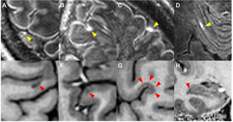
Figure 1: Leptomeningeal enhancement and cortical lesions.
Shown are examples of leptomeningeal enhancement (LME, yellow arrows) as visualized on 7T MPFLAIR and cortical lesions (red arrows) as visualized on T1-w images from 7T MP2RAGE (E – H). Four patterns of LME were noted: (A) nodular, (B) spread/fill-sulcal, (C) spread/fill-gyral, and (D) spread/fill-infratentorial. Four types of cortical lesion were also identified: (E) leukocortical, (F) intracortical, (G) subpial, and (H) hippocampal.

