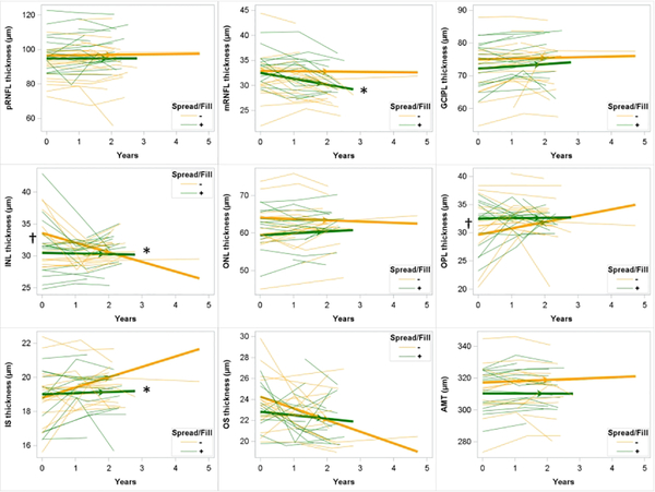
Figure 3: Longitudinal change in retinal layer thicknesses in those with and without spread/fill pattern leptomeningeal enhancement.
Spaghetti plots showing individual subject trajectories for retinal layer thicknesses. Thickened lines with arrows showing slope as determined by mixed models regression. Participants without spread/fill pattern leptomeningeal enhancement shown in yellow; those with shown in green. †: p < 0.05 for difference in baseline retinal layer thickness for those with spread/fill leptomeningeal enhancement compared to those without. *: difference in slope for those with spread/fill leptomeningeal enhancement compared to those without. LME: leptomenigneal enhancement; pRNFL: peripapillary retinal nerve fiber layer; mRNFL: macular retinal nerve fiber layer; GCIPL: ganglion cell layer + inner plexiform layer; INL: inner nuclear layer; OPL: outer plexiform layer; ONL: outer nuclear layer; IS: inner segment; OS: outer segment; AMT: average macular thickness.

