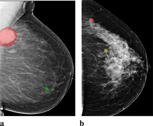
Fig. 1.

Images with both malignant and benign lesions. a. An image of the left breast from mediolateral oblique view (L-MLO). The breast has two lesions confirmed by biopsy, one as malignant (annotated with red), and the other as benign (annotated with green). b. An L-MLO mammogram image from another patient. There are two lesions on the image, one as malignant (annotated with red), and the other as high-risk benign (annotated with yellow)
