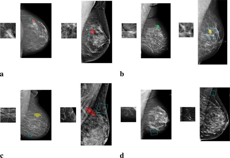
Fig. 3.
Examples of image patches along with the mammogram images from which they come. a. “malignant” patches, which overlap only with malignant findings (marked with red); b. “benign” patches, which overlap only with benign findings (marked with yellow or green); c. “outside” patches, which are from regions outside the annotated lesions; d. “negative” patches, which are from images without any biopsied findings

