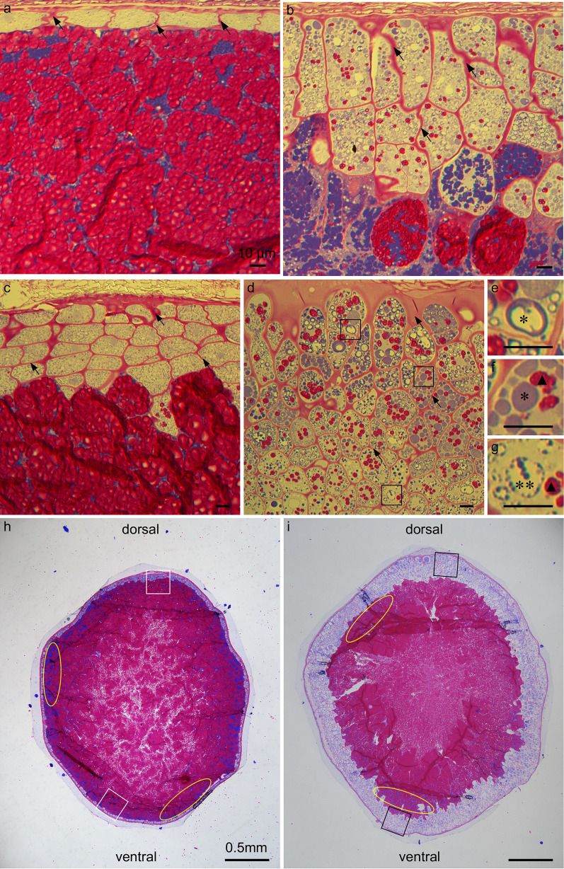
Fig. 2.
PAS and CBB staining showing the subcellular cell content of the transverse section of the rice caryopsis. PAS binds to and stains carbohydrate magenta while CBB binds to and stains protein blue. Different types of cell present across the putative multiple-aleurone layers in ta2-1. a and c the overview of the wild-type aleurone layers on the ventral side (a) and dorsal side (c) of rice caryopsis; b and d the overview of the ta2-1 aleurone layers on the ventral side (b) and dorsal side (d) of rice caryopsis; e–g the aleurone cells in the ta2-1 outer (e), middle (f), and inner (g) multiplied cell layers in three different regions of d. h and i semi-thin transverse section of the rice caryopsis showing the aleurone and starchy endosperm tissue layers in wild type (h) and ta2-1 (i). In a–d, black arrows indicate the cell wall. In d, black square indicates the locations to capture the images used in e–g. In e–g, a single asterisk indicates the protein body identified in ta2-1 resembling wild-type structure. Double asterisks indicate the large protein body in ta2-1. Black closed triangle indicates the starch granules presented in the multiple aleurone layers of ta2-1. In g, h, white square indicates the estimated locations to capture the images used in a (ventral) and b (dorsal). Black square indicates the estimated locations to capture the images used in b (ventral) and d (dorsal). Yellow oval indicates the sub-aleurone layer. Scale bar for a–g = 10 µm. Scale bar for h, i = 0.5 mm

