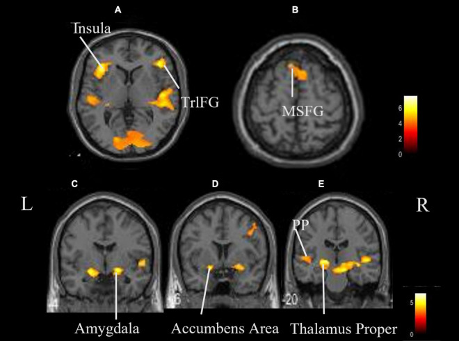
FIGURE 3.
Whole brain activation while listening to ASMR compared with the resting state. Significant activation was observed in the right triangular part of the left thalamus proper, left insula, right triangular part of the inferior frontal gyrus, right cerebellum exterior, left accumbens, right amygdala, left medial superior frontal gyrus, and left planum polare. (A,B) Axial view. (C–E) Coronal view. L, left; R, right. TrlFG, triangular part of the inferior frontal gyrus; MSFG, medial superior frontal gyrus; PP, planum polare.

