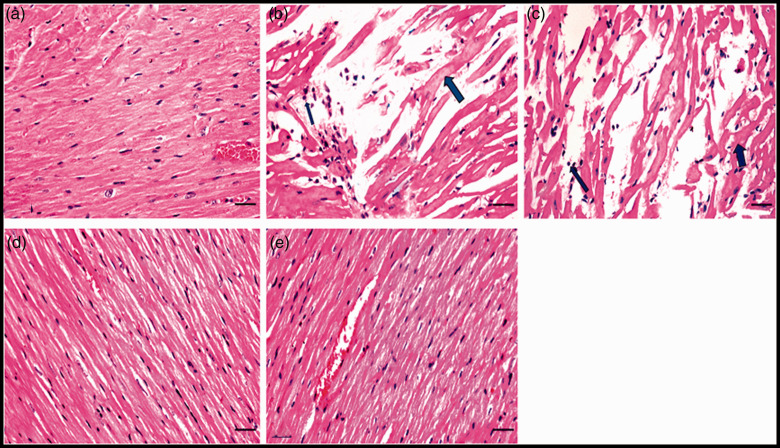
Figure 2.
Photomicrographs of cardiomyocytes in heart sections of all studied groups: (a) Heart section from negative control group showed normal myocardial muscles striation and nucleation. (b) Heart section from positive group presented myocarditis, the hyalinized myocytes (thick arrow) and leucocytic cells infiltration (thin arrow). (c) Heart section from bee venom-treated group (0.5 mg/kg) displayed hyalinized myocytes (thick arrow) and leucocytic cells infiltration (thin arrow). Histological heart section from (d) bee venom-treated group (1.23 mg/kg) and (e) metformin- and atorvastatin-treated group exhibited the normal structure of myocardial muscles (H&E staining, scale bar = 100 μM). (A color version of this figure is available in the online journal.).

