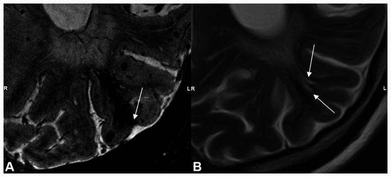
Figure 2.
Example of characteristic shape of intragyral hemorrhage and extended perivascular spaces. MRI scans of a patient with sCAA showing: (a) intragyral hemorrhage on T2*-weighted 7 T-MRI with cortical involvement (white arrow) and (b) presence of enlarged perivascular spaces (EPVS) (white arrows) in the same gyrus as the intragyral hemorrhage on T2-weighted 3 T-MRI; note the similar shape of the EPVS and the susceptibility artifact of the intragyral hemorrhage on T2-weighted MRI, possibly suggesting that intragyral hemorrhage is caused by blood leakage from a cortical microbleed into an EPVS. LR: right and left.

