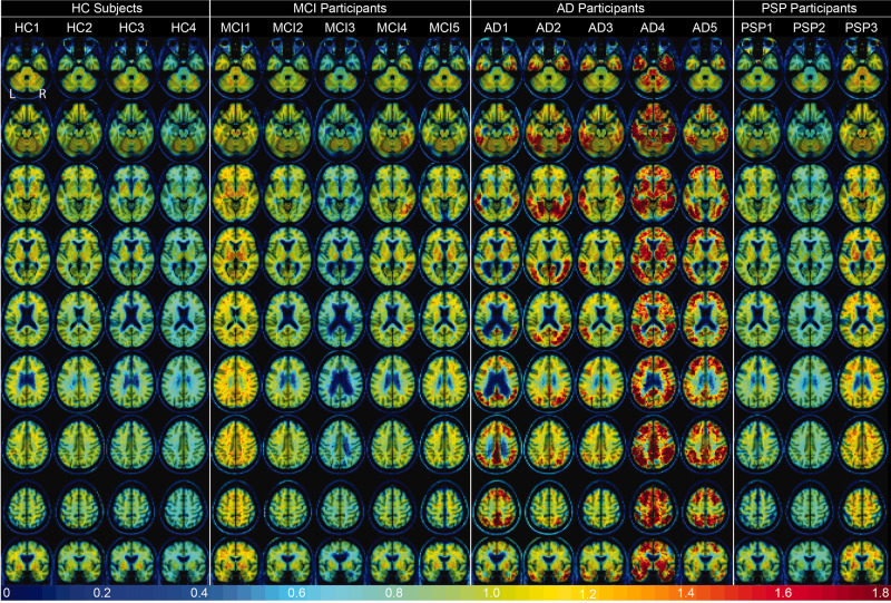
Figure 3.
[18F]-JNJ-067 DVR images (k2ref = 0.025 min−1; 35–90 min) warped to template space. Each column is a different subject following the order from Table 1, rows go through axial slices equally spaced for each subject ending with coronal showing entorhinal cortex.

