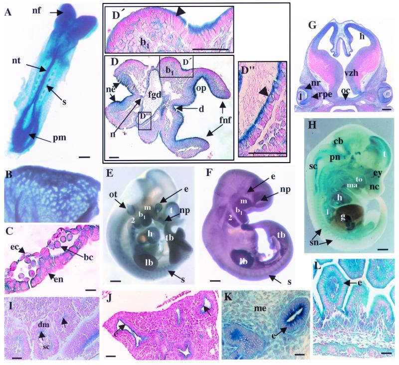
FIG. 2.
Expression of Dlg-LacZ during embryonic development in heterozygous dlggt embryos. (A) Expression in E8.5 embryos (dorsal view) was detected in the cephalic neural folds (nf), neural tube (nt), somites (s), and presomitic mesoderm (pm). Bar, 100 μm. Whole-mount LacZ staining in the vasculature of the yolk sac (E8.5) (B) and histological sections (C) demonstrated that the fusion protein was expressed in blood cells (bc), at regions of cell contact in endothelial cells (ec), and at the basal and lateral membranes of the epithelial cells of the endoderm (en). Magnification, ×100; bar, 12 μm. (D) At E8.5, transverse histological sections demonstrated that LacZ was expressed in the neural epithelium of the forebrain neural head folds (fnf), diencephelon (d), and otic pit (op), with the strongest expression detected at the apical surface. In the epithelium of the branchial arch (b1) (D′), foregut diverticulum (fgd) (D"), and surface ectoderm, expression was seen only at the apical face of the cells (arrowheads). Magnifications: panel D, ×20; panels D′ and D", ×40. Bars, 50 μm. (E) Whole-mount Dlg-LacZ expression in E10.5 embryos. (F) Endogenous Dlg expression in E10.5 wild-type embryos detected using Dlg antisera. Bars, 500 μm. (G) Dlg-LacZ expression in the brain (E12.5) was seen in the ventricular zone of the hypothalmus (vzh) and hippocampus (h). In the eye, Dlg-LacZ was expressed in the neural retina (nr), lens (l), retinal pigmented epithelium (rpe), and optic chiasm (oc). Magnification, ×5; bar, 200 μm. (H) Whole-mount Dlg-LacZ expression at E14.5 after only 4 h of X-Gal staining. Bar, 1 mm. (I) In the somites (E9.5), expression was detected in the epithelial dermomyotome (dm) component at the face adjacent to the scleratome (sc) (arrows). Magnification, ×40; bar, 25 μm. (J) Expression was seen predominantly at the apical membrane of epithelial cells (e) (arrows) within the lung (E14.5) but also at all membranes of epithelial cells of the (K) kidney (E14.5) and (L) gut (E14.5). Magnification in panel J, ×20; bar, 50 μm. Magnification in panel K, ×40; bar, 8 μm. Magnification in panel L, ×40; bar, 50 μm. b1 and b2, branchial arches 1 and 2, respectively; cb, cerebellum; ey, eye; g, gut; h, heart; lb, limb bud; l, lung; ma, mandible; m, maxillary component of b1; me, mesenchyme; nc, nasal cavity; np, nasal pit; ne, neural epithelium; n, notochord; ot, otic vesicle; pn, pons; sc, spinal cord; sn, spinal nerve; tb, tailbud; to, tongue; t, telencephalon.

