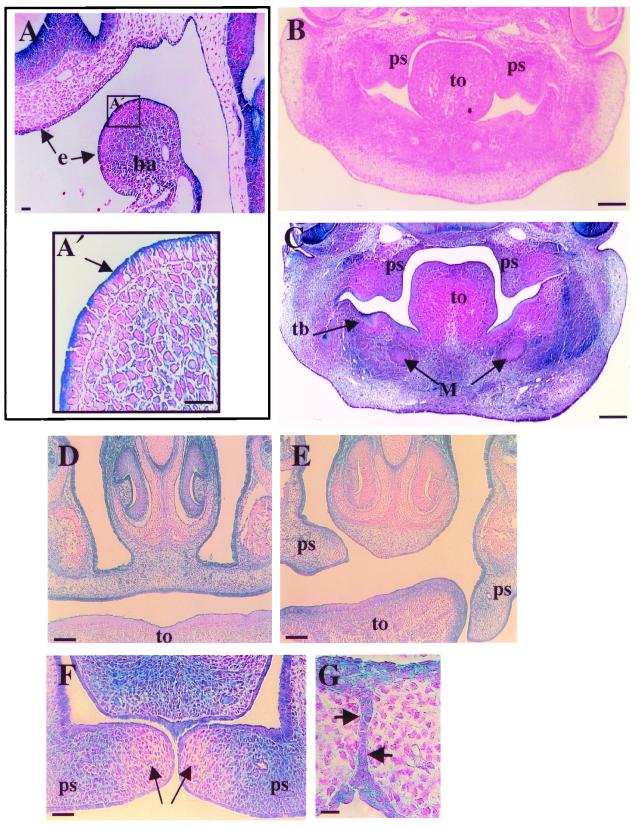
FIG. 5.
Histology of palate development and Dlg-LacZ expression in mutant dlggt embryos. (A) Dlg-LacZ was expressed at the apical membrane of the epithelium of the first branchial arch (ba) and the epithelium (e) associated with the olfactory placode in dlggt/+ embryos (E9.5) (arrows). Magnification, ×20; bar, 50 μm. (A′) Higher magnification (×40) of the boxed area in panel A demonstrating the apical expression of Dlg-LacZ in the epithelium of the branchial arch. Bar, 25 μm. Coronal sections of E12.5 wild-type (B) and dlggt/dlggt (C) heads, the latter demonstrating Dlg-LacZ expression in the palatal shelves (ps), toothbud (tb), and facial mesenchyme. Magnification, ×5; bar, 200 μm. (D) Palatal shelves are elevated and fused in dlggt/+ E15.5 embryos. (E) Only one palatal shelf is elevated in dlggt/dlggt embryos. Dlg-LacZ is expressed within the mesenchyme and epithelial cells of the fused palatal shelves (D) and unfused palatal shelves (E). Magnification, ×10; bar, 100 μm. (F) Dlg-LacZ expression in the palatal shelves of dlggt/+ prior to fusion demonstrates low levels of expression in the tips of the shelves (arrows) with higher expression adjacent to this region. Magnification, ×20. Bar, 200 μm. (G) Dlg-LacZ expression was observed in the medial-edge epithelial cells at the time of palatal-shelf fusion (arrows). Magnification, ×100; bar, 100 μm. M, Meckel's cartilage; to, tongue.

