Figure 8.
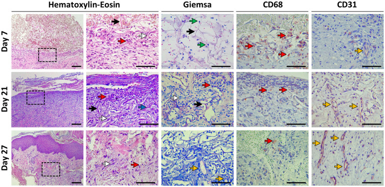
Histological evaluation. Biopsies were taken at days 7, 21, and 27 post-scaffold implantation. Histomorphology was evaluated with Hematoxylin-Eosin and Giemsa staining. Immunohistochemistry was performed to study the presence of macrophages (CD68) and endothelial cells (CD31). Colored arrows indicate: collagen fibers (black arrows), fibroblasts (white arrows), immune cells (red arrows), fibrin deposits (blue arrow), microalgae (green arrows), and vascular structures (yellow arrows). The indicated area in the left column of Hematoxylin-Eosin is magnified in the right column. Scale bars represent 100 μm for all pictures except for Hematoxylin-Eosin left column which represents 200 μm. All samples correspond to P8, except for Giemsa (P1).

