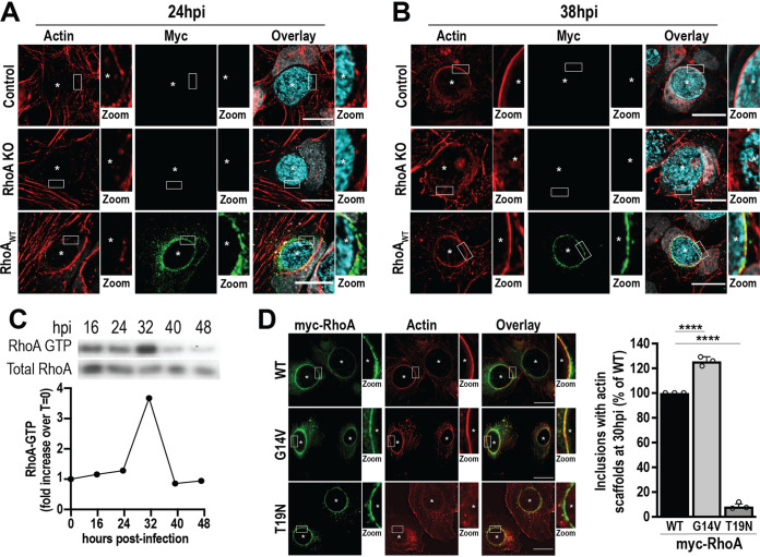
FIG 1.
Active RhoA is required for the formation of actin scaffolds around the inclusion. (A and B) Control and RhoA KO cells were infected with WT C. trachomatis L2 (MOI of 2) and transfected with empty or myc-RhoAWT DNA at 4 hpi. Cells were fixed at 24 or 38 hpi and labeled with phalloidin to label actin (red), anti-myc antibody to label RhoA (green), and anti-MOMP antibody to label individual Chlamydia (cyan). Asterisks denote inclusions. The white box indicates the magnified area shown to the right of the image (Zoom). Images are representative of 3 independent experiments. Scale bar, 25 μm. (C) HeLa cells were infected with WT C. trachomatis L2 (MOI of 1) for the indicated times prior to lysis and isolation of RhoA-GTP using the RhoA binding domain of Rhotekin immobilized on agarose beads. In the Western blot, RhoA-GTP denotes the RhoA signal from the pulldown, and Total RhoA represents the amount of RhoA from the cell lysate. The graph indicates the fold increase in RhoA-GTP compared to a noninfected control (t = 0). Results are representative of two independent experiments. (D) RhoA KO cells were infected with WT C. trachomatis L2 (MOI of 2) and transfected with myc-RhoAWT, myc-RhoAG14V, or myc-RhoAT19N DNA at 4 hpi. Cells were fixed 30 hpi and labeled with anti-myc (green) antibody and phalloidin (red). Scale bar, 20 μm. Asterisks denote inclusions. The white box indicates the magnified area shown to the right of the image (Zoom). The graph denotes the average percentage of inclusions containing actin scaffolds from 3 independent experiments ± the standard deviation. Data are normalized to myc-RhoAWT-expressing cells. A minimum of 100 inclusions was counted for each condition per experiment. ****, P < 0.0001.

