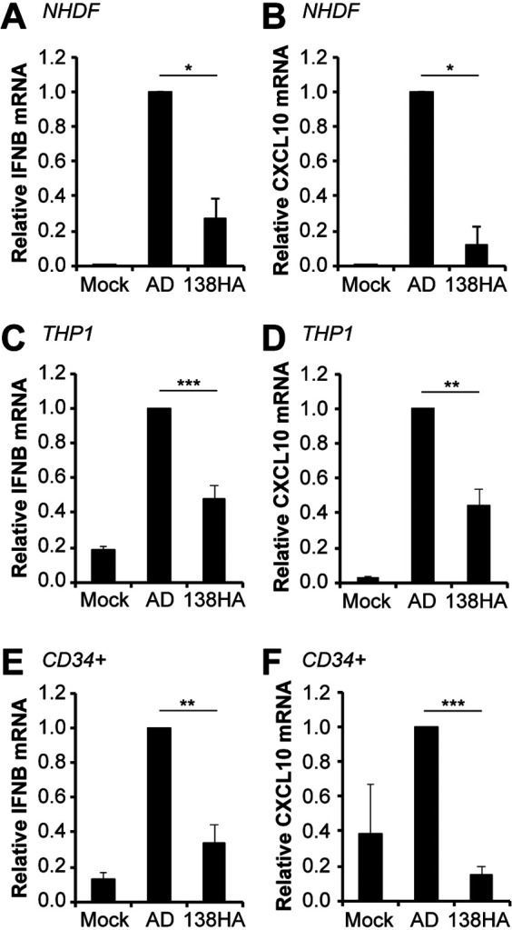
FIG 7.

A UL138-positive laboratory strain virus shows reduced IFN-β and CXCL10 transcript accumulation compared to wild-type virus during infection of either fibroblasts or myeloid cells. (A and B) NHDFs mock infected or infected with wild-type AD169 or AD169-UL138-HA virus at an MOI of 1 for 24 h were analyzed for IFN-β (A) and CXCL10 (B) transcripts by RT-qPCR. Transcript levels relative to AD169-infected cells are shown (n = 3). (C and D) THP-1 monocytes mock infected or infected with the indicated virus at an MOI of 1 for 24 h were analyzed for IFN-β (C) and CXLC10 (D) transcripts by RT-qPCR. Transcript levels relative to AD169-infected cells are shown (n = 6). (E and F) Primary CD34+ HPCs were mock infected or infected with the indicated virus at an MOI of 1 for 24 h and were analyzed for IFN-β (E) and CXCL10 (F) transcripts by RT-qPCR. Transcript levels relative to AD169-infected cells are shown (n = 5). Bar graphs show the means ± SEM from the indicated number of biological replicates.
