Figure 2.
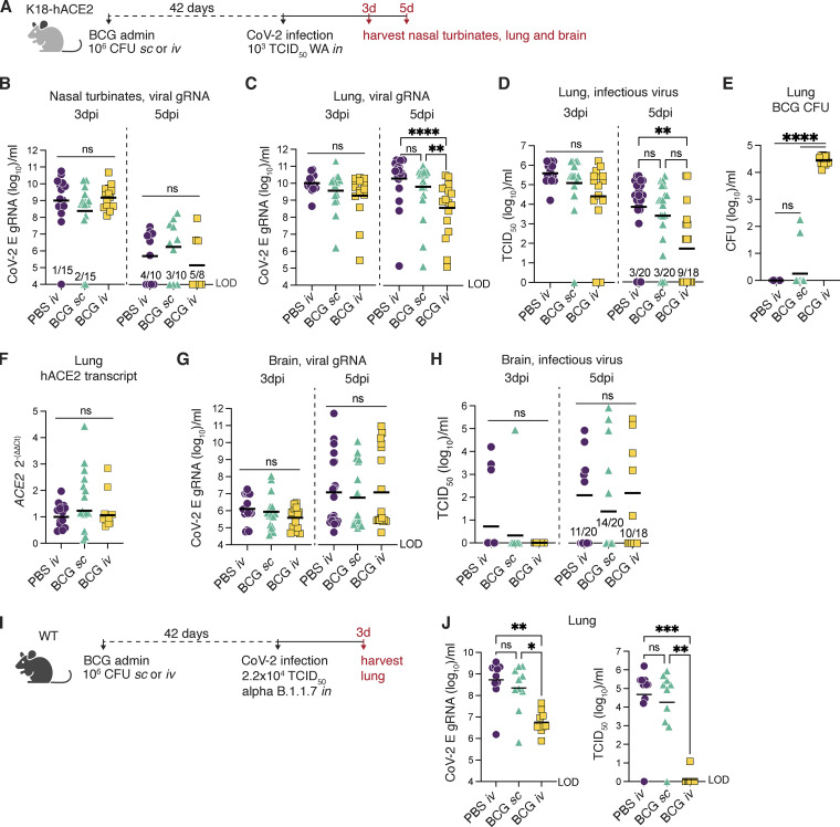
BCG i.v. administration accelerates pulmonary viral clearance in K18-hACE2 and WT mice. (A–H) K18-hACE2 mice were inoculated with 106 CFUs BCG Pasteur by s.c. or i.v. injection. Control animals received the same volume of PBS i.v. At 42 d after BCG administration, mice were infected with 103 TCID50 SCV2 (WA1/2020) by i.n. instillation. (B) Nasal turbinates were collected 3 or 5 d after challenge and assessed for viral load by qPCR. (C–F) Lungs were collected 3 or 5 d after viral challenge and assessed for viral load by qPCR and TCID50 assay (C and D), mycobacterial load by counting CFUs (E), and expression of human ACE2 by RT-qPCR (F). (G and H) Viral loads were quantified in brain samples at 3 and 5 dpi by qPCR and TCID50 assay. (I and J) C57BL/6 (WT) mice were inoculated with 106 CFUs BCG Pasteur by s.c. or i.v. injection. Control animals received the same volume of PBS i.v. At 42 d after BCG inoculation, mice were infected with 2.2 × 10⁴ TCID50 SCV2 (α B.1.1.7) by i.n. instillation. Lungs were collected 3 d after viral challenge, and homogenates were assessed for viral load by qPCR and TCID50 assay (J). Statistical significance was assessed by Kruskal-Wallis test with Dunn’s post-test. ns, P > 0.05; *, P < 0.05; **, P < 0.01; ***, P < 0.001; ****, P < 0.0001. Data are pooled from two to four independent experiments each with 4–10 mice per group. Graphs show geometric mean and individual values for each animal. LOD, limit of detection.

