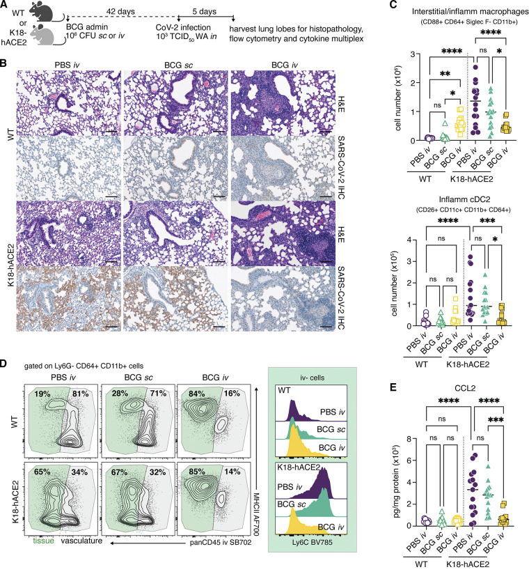
Figure 3.
SCV2-driven inflammatory cellular responses are dampened in mice previously inoculated with BCG i.v. (A–E) K18-hACE2 mice or non-transgenic littermate controls (WT) were inoculated with 106 CFUs BCG Pasteur by s.c. or i.v. injection. Control animals received the same volume of PBS i.v. At 42 d after BCG administration, mice were infected with 103 TCID50 SCV2 (WA1/2020) by i.n. instillation. (A) Lungs were collected 5 d after viral challenge and assessed by histopathology, flow cytometry, and cytokine multiplexing. (B) Representative images of H&E-stained lung tissue sections (top panels) and sequential sections probed with an anti–CoV-2 nucleoprotein antibody (bottom panels). Scale bars: 100 μm. (C) Number of interstitial/inflammatory macrophages and inflammatory cDC2. (D) Contour plots depict MHCII expression by tissue-resident (i.v. stain negative; green) and vascular (i.v. stain positive; gray) CD64+ CD11b+ cells in the lung (left). Histograms show Ly6C expression by i.v.− CD64+ CD11b+ cells (right). Data are concatenated from four to five mice per group and are representative of three independent experiments. (E) CCL2 levels in lung tissue homogenate standardized to total protein concentration. Statistical significance was assessed by one-way ANOVA with Tukey post hoc test. ns, P > 0.05; *, P < 0.05; **, P < 0.01; ***, P < 0.001; ****, P < 0.0001. Unless otherwise stated, data are pooled from three independent experiments each with four to five mice per group. Flow cytometry gating strategy is shown in Fig. S2. IHC, immunohistochemistry.

