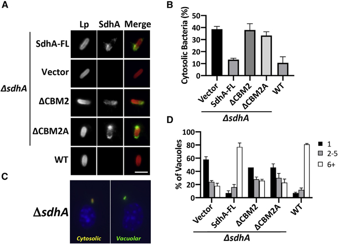
Figure 3. The OCRL binding region of SdhA is required for maintaining LCV integrity and intracellular growth in macrophages.
BMDMs from the A/J mouse were challenged at MOI = 1 with L. pneumophila WT or ΔsdhA harboring SdhA variants.
(A) SdhA localization on LCV was determined by immunofluorescence using antibodies against SdhA at 3 hpi (scale bar, 2 μm). See also Figure S1.
(B) SdhA mutants do not rescue ΔsdhA vacuole disruption. Macrophages were challenged for 8 h, fixed, and stained for bacteria before and after permeabilization, and internalized bacteria in absence of permeabilization were quantified relative to total infected population (mean ± SD; n = 3).
(C) Examples of cytosolic and vacuolar bacteria. Macrophages were challenged with the ΔsdhA strain, fixed, probed with α-L. pneumophila (Alexa Fluor 594 secondary, red), permeabilized, and reprobed with α-L. pneumophila (Alexa 488 secondary, green; STAR Methods). Cytosolic bacteria are accessible to both antibodies (yellow).
(D) Growth of L. pneumophila strains in macrophages, determined by the number of bacteria per vacuole 16 h post-uptake.
(B and D) Data represent mean ± SD of biological triplicates. More than 100 LCVs were counted per replicate. Linked to Figure S1.

