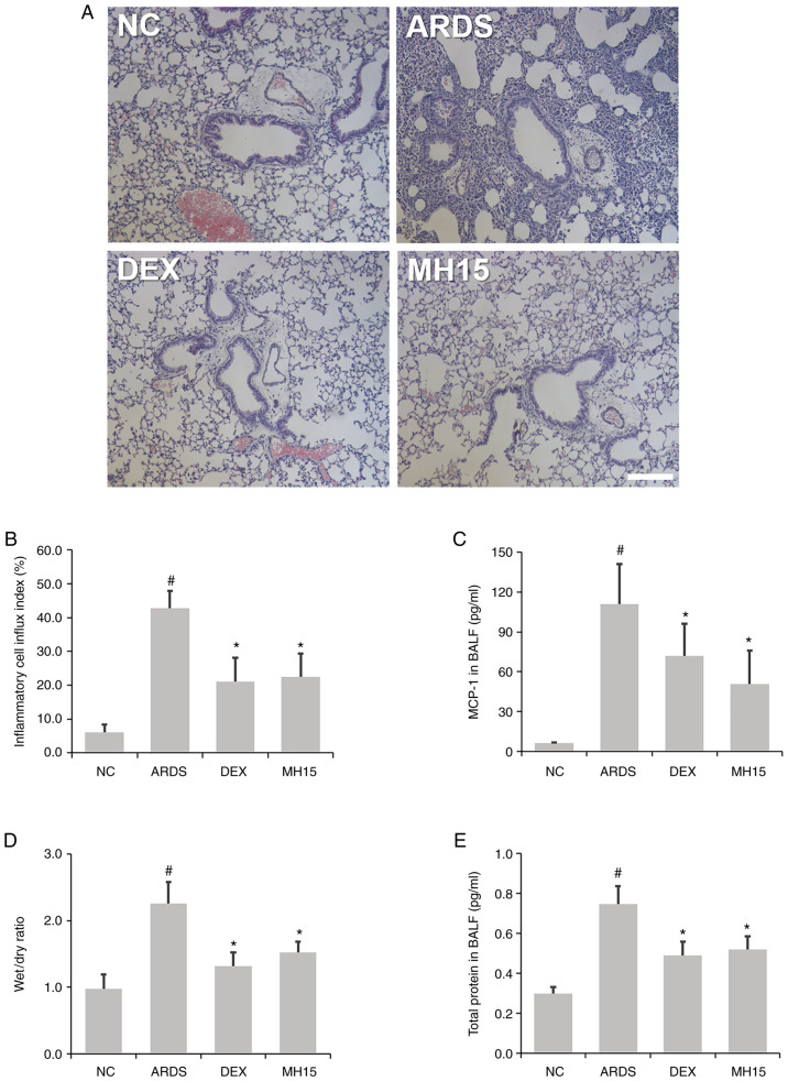
Figure 4.
MH suppresses inflammatory cell influx and MCP-1 secretion in lipopolysaccharide-induced ARDS mice. (A) Hematoxylin and eosin staining was used to detect inflammatory cells around the airways in the lungs of mice (magnification, ×100; scale bar, 100 µM). (B) Quantitative analysis of airway inflammation. (C) ELISA was used to determine the secretion level of MCP-1 in BALF of mice. (D) Lung wet/dry ratio and (E) BALF protein were evaluated to determine lung edema. Data are expressed as the mean ± SD. #P<0.05 vs. NC; *P<0.05 vs. ARDS. MCP-1, monocyte chemoattractant proein-1; NC, normal control; ARDS, acute respiratory distress syndrome; DEX, dexamethasone; MH, methyl p-hydroxycinnamate; BALF, bronchoalveolar lavage fluid.

