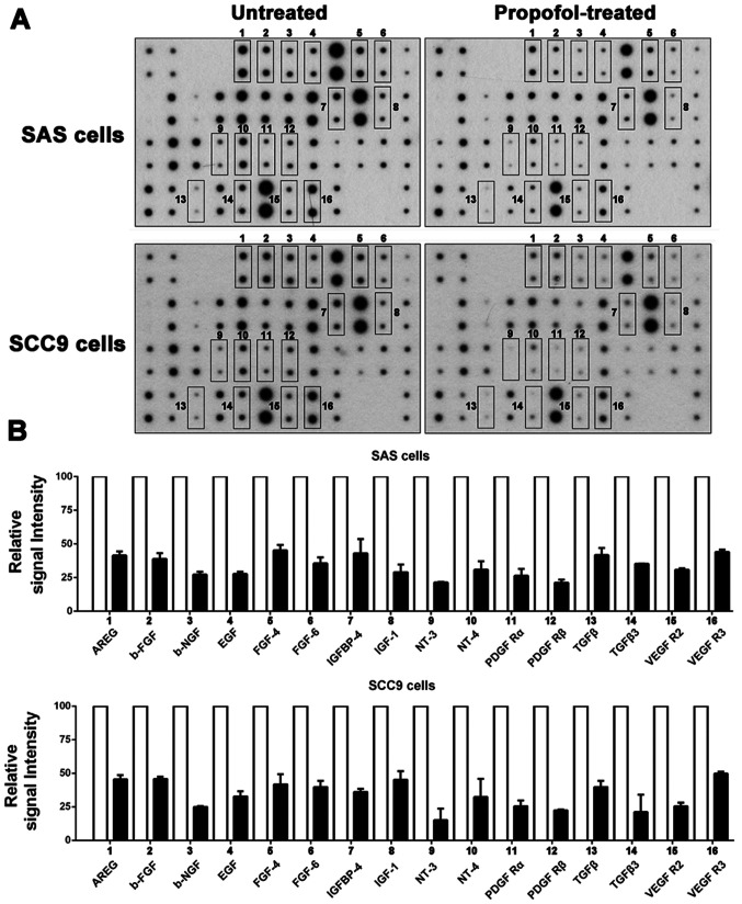
Figure 2.
Effect of propofol on protein secretion in oral squamous cell carcinoma cells. (A) Cells were treated with 10 µM propofol for 48 h. Untreated cells were used as controls. Serum-free conditioned media was harvested and analyzed using a human growth factor antibody array. Each antibody was spotted in duplicate onto a membrane. The boxes with numbers indicate that the spots that were markedly reduced in size in the propofol-treated group compared with the untreated group (fold changes >2). (B) With reference to untreated cells, the intensity of the spots was quantified by densitometry analysis and displayed as a relative signal intensity. AREG, amphiregulin; b-FGF, basic fibroblast growth factor; b-NGF, beta-nerve growth factor; IGFBP, insulin like growth factor binding protein; IGF, insulin like growth factor; NT, neurotrophin; PDGF R, platelet derived growth factor receptor.

