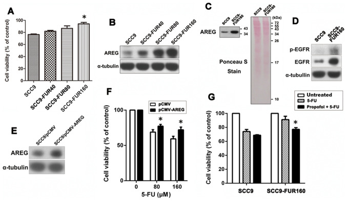
Figure 5.
AREG upregulation is related to the development of 5-FU resistance, but propofol alleviates 5-FU resistance. (A) Cells were treated with 160 µM 5-FU for 48 h and then cell viability was determined by performing MTT assays. Data are presented as the mean ± SEM (n=3). *P<0.05 vs. SCC9. (B) AREG protein expression levels in 5-FU-resistant cell sub-lines and their parental cells were analyzed by western blotting with α-tubulin as the loading control (n=3). (C) Secreted levels of AREG in the cell culture media of SCC9 and SCC9-FUR160 cells were analyzed using western blotting (n=3). Ponceau S staining demonstrated that equivalent amounts of total protein were loaded on each lane. (D) Basal expression of p-EGFR and total EGFR in SCC9 and SCC9-FUR160 cells was evaluated by western blotting (n=3). (E) SCC9 cells were transiently transfected with pCMV3-AREG and then the protein expression level of AREG was analyzed by western blotting (n=3). (F) Cells were treated with increasing concentrations of propofol (0–160 µM) for 72 h. Effect of AREG overexpression on 5-FU-induced cytotoxicity was determined by performing MTT assays (n=4). *P<0.05 vs. pCMV. (G) Cells were treated with propofol (10 µM) for 24 h and then stimulated with 5-FU (40 µM) in the presence of 10 µM propofol for 48 h. Cell viability was then assessed by performing MTT assays (n=3). *P<0.05 vs. 5-FU treatment alone. AREG, amphiregulin; 5-FU, 5-fluorouracil; p, phosphorylated; FUR, 5-FU-resistant. F, 5-FU.

