
Fig 5. U. parvum promotes EMT in endocervical epithelial cells.
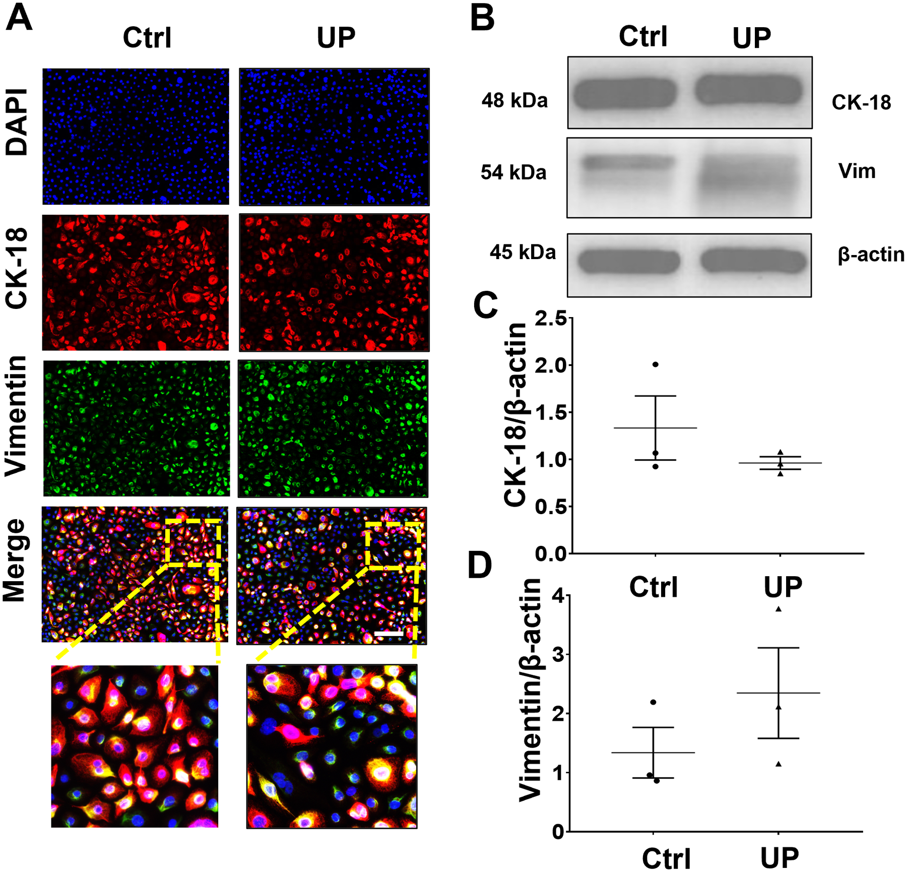
(A) Fluorescence microscopy imaging showing CK-18 and vimentin in uninfected and U. parvum-infected human endocervical epithelial cells. Nuclei are stained with DAPI, n=3 technical replicates. Scale bar, 100 μm. Western blot analysis and quantification of CK-18 (B, C) and vimentin (B, D) in endocervical epithelial cells. β-actin is a loading control. Error bars represent mean ± SEM, n=3 technical replicates. Linear adjustment of contrast and brightness has been applied to all fluorescent images throughout the figure.