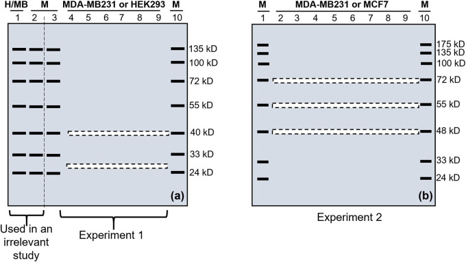
Figure 1.
Illustration of excision of narrow stripes of gel after SDS-PAGE in two (a and b) experiments. (a) Two gels were made for the first experiment. The first well was loaded with proteins from HEK293 cells (H) in one gel and proteins from MDA-MB231 cells (MB) in the other gel. The second, third, and tenth wells of both gels were loaded with a prestained protein marker (M). The fourth to ninth wells of one gel were loaded with proteins from HEK293 cells, but these wells of the other gel were loaded with proteins from MDA-MB231 cells. After electrophoresis, both gels were cut into two parts along the vertical dashed line between the second and third lanes. The left part of both gels containing lanes 1 and 2 was used in WB to detect the CDK4 protein isoforms at the 40 and 26 kD positions, which was the initial purpose of this experiment but is irrelevant to the present study. The right part of both gels was used for this study, of which two narrow stripes (illustrated as dashed boxes) were excised at the 26 and 40 kD positions. (b) Two gels were also made in the second experiment. Although the first and the last wells of both gels were loaded with a prestained protein marker (M), the remaining wells were loaded with proteins from MDA-MB231 cells in one gel and proteins from MCF7 cells in the other gel. After electrophoresis, three narrow stripes shown as dashed boxes were excised from each gel at the 72, 55, and 48 kDa positions. All ten stripes from all the four gels of these two experiments were later used for LC-MS/MS analyses.

