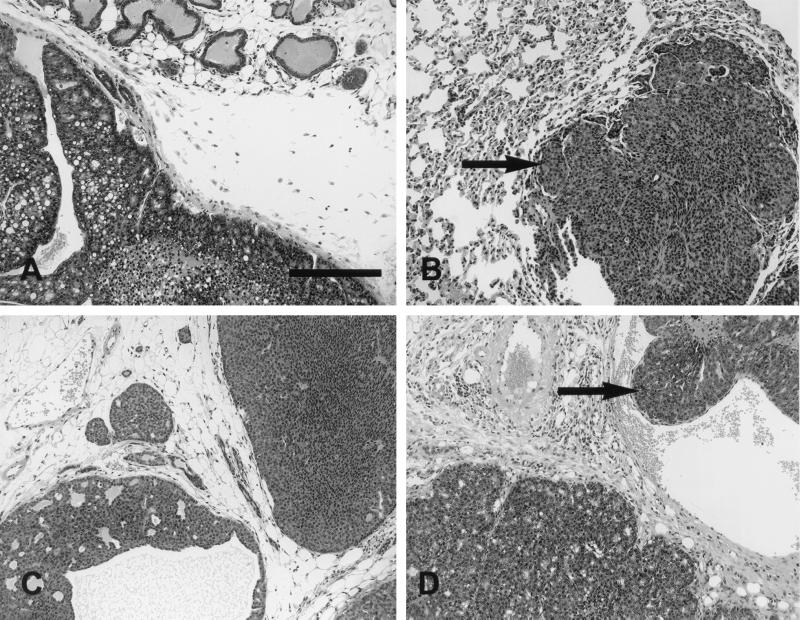
FIG. 7.
Histopathology of mammary tumors induced by the various neu autophosphorylation mutants. Photoimages show mammary tumors from mice expressing the neu autophosphorylation mutants NYPD (line 10) (A and B), YD (line 6) (C), and YB (line 2) (D). All three animals exhibited well-differentiated glandular patterns. The tumors were composed of cells with relatively small, oval to round nuclei without significant pleomorphism. The cells were cytologically identical to the cells seen in all Neu-induced tumors. YB consistently had a better-differentiated glandular pattern (D). The size bar indicates 0.1 mm.

