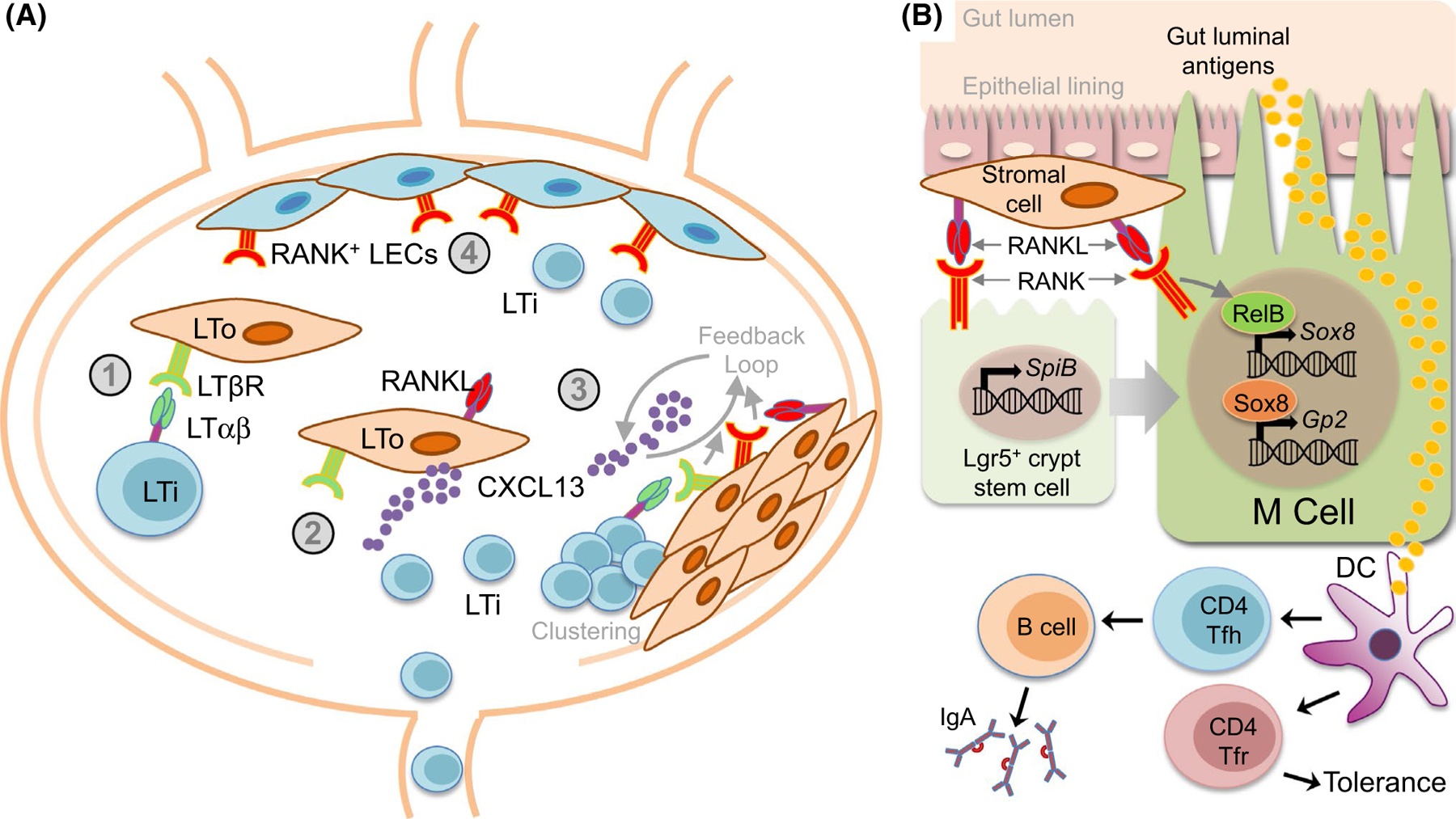
Fig. 2.

RANKL-RANK-OPG contributes to secondary lymphoid organogenesis. a RANKL-RANK-OPG contributions to lymph node organogenesis. Initiation of lymph node (LN) formation involves recruitment of hematopoietic lineage lymphoid tissue inducer (LTi) cells to LN anlagen that include lymphoid tissue organizer (LTo) progenitor/precursor cells. (1) LTi cells expressing RANKL, RANK, and LTαβ interact with LTβR-expressing LTo cells. (2) Activation of LTβR induces LTo cells to mature, increasing expression of RANKL and the chemokine CXCL13, which increases attraction of more LTi cells. (3) LTi cells then cluster with the LTo cells to initiate LN formation. Since LTo cells may also express RANK, a positive feedback loop develops involving LTo and LTi cells with RANKL-RANK and LTαβ-LTβR signaling amplifying the recruitment-clustering process of lymph node development. (4) RANK-expressing lymphatic endothelial cells (LECs) line the LN subcapsular sinus and act as another type of LTo that are critical to LTi cell retention, via RANKL-RANK interactions, during LN organogenesis. b Peyer’s patch microfold (M) cells. Peyer’s patches (PP) are a gut-associated lymphoid tissue (GALT) found along the small intestine where CD4+ follicular helper T cells (Tfh) are critical components of germinal centers necessary for gut-associated IgA responses. CD4+ follicular regulatory T cells (Tfr) also inhabit PPs to regulate peripheral tolerance. Microfold (M) cells are specialized epithelial cells that transport gut luminal antigens for presentation to PP T cells. RANK signaling is required for M cell differentiation and antigen uptake. RANKL expression by subepithelial stromal cells in PP domes drive M cell differentiation. RANK signaling contributes to the earliest stages of M cell differentiation by inducing expression of the transcription factor SpiB in M cell precursor Lgr5+ intestinal crypt stem cells. RANK-dependent activation of the NF-κB factor RelB is required for expression of the transcription factor Sox8, which then activates the promoter of the key M cell factor Gp2
