Fig. 6.
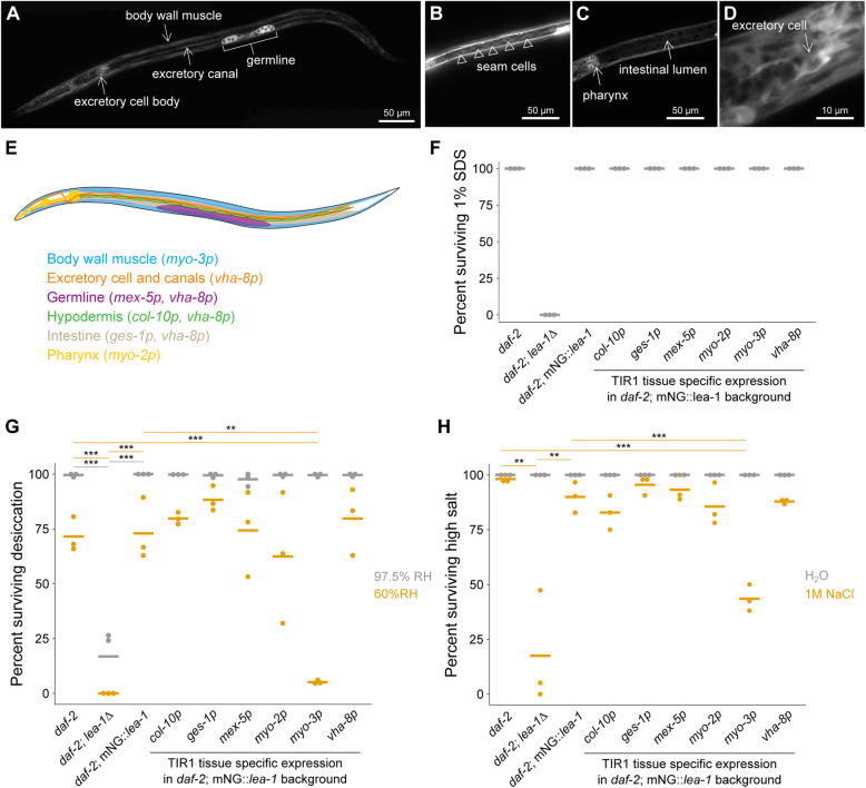
LEA-1 is required in body wall muscle to survive desiccation and osmotic stress. A–D Representative images of an mNG::lea-1 worm depict the major sites of expression. There is prominent fluorescence in the germline, body wall muscle, excretory cell, and pharynx. There is also some apparent expression in seam cells (B) and faint fluorescence in the intestine (C). A zoomed in image of the excretory cell is shown in D. E A cartoon depicts tissues in which LEA-1 is expressed that were targeted for protein depletion by driving TIR1 under the control of different promoters. F Survival in 1% SDS is plotted for worms with tissue specific LEA-1 depletion by expression of TIR1 under various promoters and exposure to 1 mM auxin. Dauer larvae were formed at 25 °C. Depletion of LEA-1 did not sensitize worms to SDS. G Desiccation survival is plotted for worms with tissue-specific LEA-1. Depletion of the mNG::lea-1 protein utilizing auxin-induced degradation reduces desiccation survival in worms expressing TIR1 under a myo-3 (body wall muscle-specific) promoter relative to daf-2 controls (p = 0.0001, n = 3, unpaired T test) and worms expressing only mNG::lea-1 and not TIR1 (p = 0.001, n = 3, unpaired T test). No other site of TIR1 expression significantly altered desiccation survival at 60% RH. H Survival of osmotic stress in 1 M NaCl for 4 h is plotted for the same strains as in (E). lea-1Δ mutants and worms expressing TIR1 under a myo-3 promoter were the only two strains that with significant differences in survival relative to both control and mNG::lea-1-expressing animals (daf-2;lea-1Δ: vs. daf-2 p = 0.006, vs. daf-2;mNG::lea-1 p = 0.01; daf-2;mNG::lea-1;myo-3p::TIR1: vs. daf-2 p = 0.0001, vs. daf-2;mNG::lea-1 p = 0.0009, unpaired T tests, n = 3). Note that the images in A–D show a worm with an N2 background, whereas worms in F–H are in a daf-2(e1370) background. **p < 0.01, ***p < 0.001

