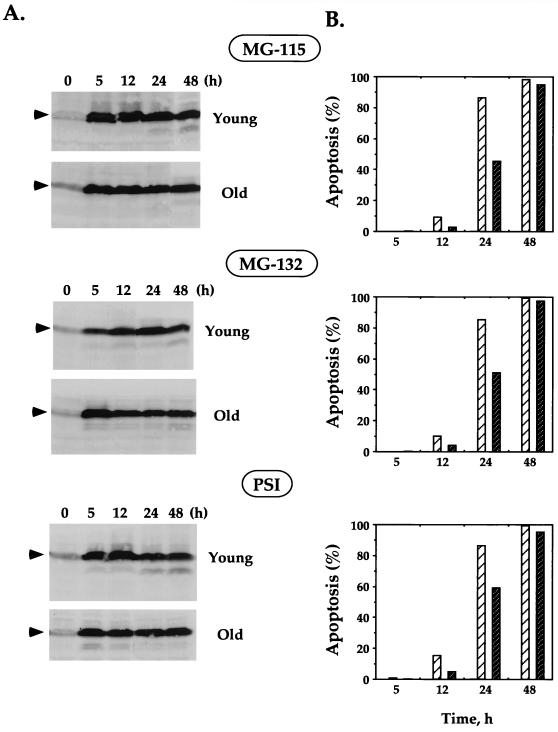
FIG. 6.
Stabilization of p53 and induction of apoptosis in young and old fibroblasts treated with proteasome inhibitors. Young and old fibroblasts were treated with the following proteasome inhibitors: MG-115 (30 μM), MG-132 (10 μM), and PSI (30 μM). (A) Stabilization of p53 at various time points after treatment was analyzed by Western blotting with DO-1 antibodies. The p53 protein is indicated by arrowheads. The blots were stained with India ink to check the equivalence of protein loading and transfer, and the blots showing equal loading and transfer within young and old cells are presented. (B) Induction of apoptosis was analyzed by acridine orange staining followed by FACS analysis. The percent apoptosis in young (light hatched bars) and old (dark hatched bars) cells treated with proteasome inhibitors is shown. The level of apoptosis in untreated cells (young and old) was too low to be seen on the graph. Experiments were repeated at least three times; standard errors were less than 3% of the average and are not shown on the graph.

