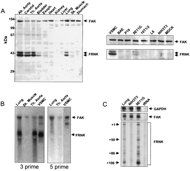
FIG. 1.
Expression of FRNK in smooth muscle tissues and cultured cells. (A) Individual tissues from rats (4-day-old pups) were dissected, washed in PBS, and lysed in RIPA buffer. Protein extracts (100 μg/lane) from tissues (left) or cultured cells (right) were subjected to SDS-PAGE. Western blotting was performed using an anti-FAK carboxyl-terminal Ab. (B) RNA prepared from rat tissues (3-week-old pups) or vascular SMCs was subjected to Northern analysis using a 3′ or 5′ cDNA probe from the FAK coding region (corresponding to nucleotides 2296 to 3213 or 1163 to 2088, respectively). The arrows indicate the positions of the FAK and FRNK RNAs. (C) A sequencing gel showing the positions of labeled, RNase-resistant products obtained after incubation of total mouse lung, 9E11G RNA, or NIH 3T3 RNA with a probe for glyceraldehyde-3-phosphate dehydrogenase (GAPDH), FAK, or FRNK. Abbreviations: Ab., abdominal; Th., thoracic; Sk., skeletal; VSMC, rat aortic SMC; BAE, bovine aortic endothelial cells.

