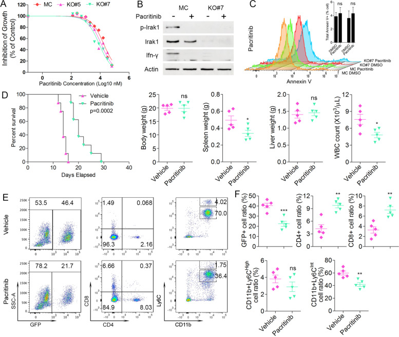
Fig. 8.
Treatment of MC BBC2 cells and IRAK1 KO #5 and #7 cells (N = 3) with pacritinib shows no differential effects on cell growth inhibition at high doses as determined using CellTiter-Glo Cell Viability Assays (A). Pacritinib treatment of the MC cells which express IRAK1 leads to suppression of its phosphactivation and loss of IFN-γ production (B). Flow cytometry analysis of annexin V staining (N = 3) of the same cells from (B) shows no differential induction of apoptosis in either the MC of KO Cells (C). When BBC2 cells were xenografted into BALB/c mice (N = 8) and then treated with the pacritinib IRAK1 inhibitor, there was a significant increase in survival time (D), which was reflected in a significantly reduced spleen size and reduction in white blood cell count in the treated cohort. Representative flow cytometry analysis of peripheral blood from the treated and control cells (E) at the time of sacrifice shows reduced levels of GFP+ cells in the drug-treated mice (N = 5) and a significantly higher level of CD4+/CD8+ T-cells (F). Levels of MDSC in the drug treated mice was also reduced compared with mice treated with vehicle alone. ns = not significant, * p < 0.05, ** p < 0.01, *** p < 0.001

